Osteoarthritis Physiotherapy: Managing Pain and Improving Movement
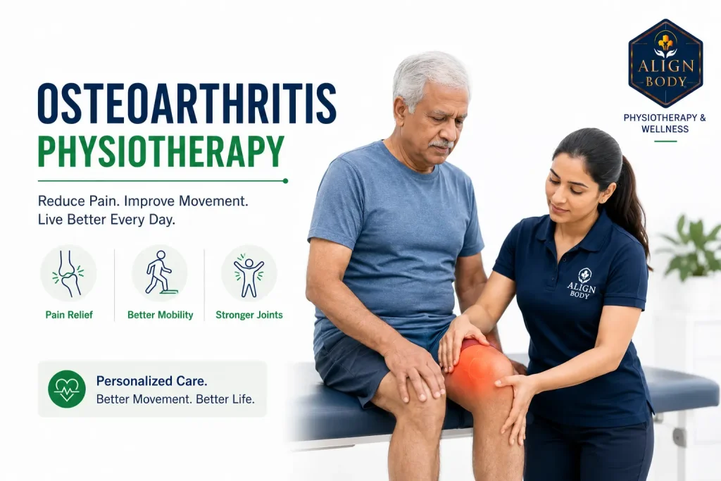
Osteoarthritis (OA) is now understood as a whole-joint condition involving cartilage, bone, synovium and surrounding muscles, not simply “wear and tear” from overuse. Exercise-based physiotherapy is genuinely first-line treatment in international clinical guidelines, ahead of medications and injections. Most patients with mild to moderate knee or hip OA experience meaningful pain reduction and functional improvement with structured physiotherapy over 3 to 6 months. Surgery becomes a consideration when conservative treatment has been properly tried and quality of life remains significantly affected. Delay in starting physiotherapy is one of the biggest predictors of eventual need for surgery.
Osteoarthritis is one of the most common conditions we treat and one of the most frequently misunderstood. Patients arrive believing their joint is “worn out” and their only option is eventual replacement surgery. They have been told to “avoid activity” to prevent further damage. They have accepted the pain as an inevitable part of ageing. Every one of these beliefs is wrong or, at best, incomplete.
Current research has fundamentally changed how OA is understood and treated. It is no longer viewed as a simple mechanical wear-out of cartilage but as a whole-joint condition involving inflammation, metabolic factors, muscle weakness and biomechanical loading patterns that can be modified. This shift matters because it changes what actually works. Exercise, once feared as potentially damaging, is now the strongest evidence-based first-line treatment.
I am Dr. Richa Gupta, founder of AlignBody Physiotherapy Clinic in Delhi. This post covers what OA actually is (versus the wear-and-tear myth), why exercise protects rather than damages OA joints, joint-specific approaches for knee, hip and hand OA, honest evidence on injection therapies, when surgery becomes appropriate and how to manage pain flares without derailing progress.
What Osteoarthritis Actually Is
The old model of osteoarthritis was mechanical: cartilage wears down from decades of use, the joint deteriorates and eventually needs replacement. Current understanding is different and more useful for treatment.
Two ways of understanding OA. Only one leads to good treatment:
✗ Old model (misleading)
Wear and tear from overuse
- Cartilage mechanically wears out
- More activity = more damage
- Rest and avoid loading
- Wait for surgery when bad enough
✓ Current understanding
Whole-joint metabolic condition
- Cartilage, bone, synovium, muscles all involved
- Inflammatory + metabolic factors matter
- Movement protects cartilage nutrition
- Exercise strengthens supporting muscles
The old model discourages exercise. The current model shows exercise is the strongest first-line treatment.
OA involves the whole joint, not just cartilage. The tissues affected include:
- Cartilage: loss of proteoglycans, disorganisation of collagen structure, thinning over time
- Subchondral bone: remodelling changes, bone marrow lesions that correlate with pain, subchondral sclerosis
- Synovium: low-grade inflammation, thickening of the joint lining
- Ligaments and joint capsule: structural changes, sometimes laxity or tightening
- Muscles surrounding the joint: weakness, altered activation patterns, atrophy
- Meniscus (in knee OA): degenerative changes, small tears often coexist
What produces pain in OA is not the amount of cartilage loss on an X-ray. Many patients with severe cartilage loss have minimal pain. Many patients with only moderate changes on imaging have significant symptoms. The relationship between X-ray findings and pain experience is weaker than most patients realise. This is why treatment that changes muscle strength, movement patterns, joint loading and inflammatory factors often produces meaningful pain relief even when the underlying cartilage change is not itself reversed.
The Exercise Paradox: Why Movement Protects Rather Than Damages
A common belief among OA patients is that exercise will accelerate joint damage. The opposite is closer to the truth. Understanding why matters for engaging with treatment.
Cartilage does not have a direct blood supply. It receives nutrition through joint fluid, which is circulated by the compression and release cycles of normal joint movement. Immobility literally starves cartilage. Regular varied loading is what maintains cartilage health, within reasonable limits.
Additionally, the muscles around the joint are the main shock absorbers during activity. Weakened muscles from disuse mean each step transmits more force directly through the joint. Strengthening surrounding muscles offloads the joint. A patient with strong quadriceps and gluteal muscles transmits less peak force through an osteoarthritic knee with every step than the same patient with weakened muscles.
The evidence is now consistent across dozens of systematic reviews and international clinical guidelines. Exercise-based physiotherapy is first-line treatment for knee and hip OA, ahead of medications and injections. This is not a preference or a philosophical position; it is what the evidence supports and what current guidelines recommend.
What this looks like practically: progressive strengthening of the muscles supporting the affected joint, aerobic conditioning to reduce systemic inflammation and maintain function, movement pattern correction to reduce peak joint loading and, in some cases, aquatic exercise for patients who cannot tolerate weight-bearing exercise on land. Read our detailed guide on kinesiotherapy for chronic pain for more on the exercise mechanisms.
Knee OA: The Specific Approach
Knee OA is the most common form we see in clinical practice, particularly in patients over 50. It typically presents as gradual-onset pain with weight-bearing activities, stiffness after prolonged sitting (the “movie theatre sign”), morning stiffness lasting under 30 minutes and reduced walking distance.
Effective treatment includes:
- Quadriceps strengthening: the single most important intervention for knee OA. Weak quads are consistently associated with worse knee OA outcomes
- Gluteal strengthening: weak glutes cause abnormal knee loading patterns during walking and stair climbing
- Progressive aerobic conditioning: walking, cycling and swimming all have evidence. Choose what the patient can sustain
- Weight management where relevant: each kilogram of body weight lost reduces knee joint load approximately 4 kilograms with every step
- Neuromuscular exercise (GLA:D approach): a specific programme with strong evidence for knee OA involving balance, control and functional movement work
- Manual therapy: joint mobilisation and soft tissue work as adjuncts, not as primary treatment
- Footwear and orthotic advice where relevant to reduce joint loading
Hip OA: What Differs
Hip OA often presents differently from knee OA. Pain typically sits in the groin or front of hip rather than the hip itself, morning stiffness may be longer, difficulty putting on socks or shoes is characteristic and walking distance is often more limited than the imaging findings would suggest. Read our detailed guide on hip pain physiotherapy for the full differential.
Treatment adapts to the hip’s specific mechanics:
- Gluteal strengthening (particularly gluteus medius) is even more important than in knee OA
- Hip flexor mobility work matters because sustained sitting often causes secondary hip flexor tightness
- Core strengthening supports the whole pelvic-hip complex
- Manual therapy of the hip joint capsule can be particularly useful in hip OA
- Aquatic exercise is often especially beneficial because unloading the joint allows patients to work through greater range of motion
Hand OA and Thumb Base OA
Hand OA is common but often undertreated. It typically affects the thumb base (first carpometacarpal joint) and the distal finger joints, producing Heberden’s nodes (visible bony enlargements at the finger-tip joints) and Bouchard’s nodes (at the middle finger joints).
Treatment approach:
- Grip and pinch strengthening (progressive, avoiding pain flares)
- Splinting for thumb base OA during high-demand activities
- Joint protection strategies (larger tool handles, adaptive equipment for jars and door handles)
- Heat therapy for symptomatic relief before activity
- Manual therapy for stiff joint capsules
Hand OA is often more visibly noticeable than functionally limiting. Reassurance about this is often part of treatment.
The GLA:D Programme: Evidence-Based Structured OA Care
GLA:D (Good Life with osteoArthritis: Denmark) is a specific evidence-based programme developed in Denmark for knee and hip OA. It involves two sessions of patient education followed by 12 supervised neuromuscular exercise sessions over 6 to 8 weeks. Multiple studies from Denmark and other countries have shown meaningful pain reduction (around 30 percent), improved function, reduced medication use and reduced surgery consideration in participants.
The GLA:D approach is not the only evidence-based programme, but it exemplifies the current standard: structured, progressive, individualised, combining exercise with education. Delhi clinics offering “OA exercises” without this structured approach are typically delivering less effective care.
Whether or not the specific GLA:D branding is used, look for these features in an OA programme: initial detailed assessment, education about what OA actually is (not wear-and-tear framing), progressive supervised exercise over at least 6 weeks, muscle strengthening plus neuromuscular control training, home exercise programme, follow-up review at intervals.
The evidence-based OA treatment ladder:
First-line (guidelines): Exercise + education + weight management
6-12 weeks of structured supervised progressive exercise. Should be tried before injections or surgery.
Adjuncts: Topical NSAIDs, paracetamol, manual therapy
Short-term flare management. Never replaces exercise. Topical NSAIDs preferred over oral for older adults.
Injection therapies (selective use)
Cortisone for acute flares (max 2-3/year). HA and PRP as adjuncts, not replacements. Always alongside exercise.
Joint replacement surgery
Only after 3-6 months of proper structured physiotherapy has been tried. Excellent outcomes when correctly indicated.
Injection Therapies: What the Evidence Actually Shows
Patients often arrive having been offered various injection treatments. Here is what current evidence suggests for each.
Cortisone (corticosteroid) injections
Cortisone injections into an OA joint reduce pain moderately for 2 to 6 weeks in most patients. Beyond that, effect diminishes. Repeated cortisone injections (more than 2 to 3 per year) have been associated with faster cartilage loss and worse long-term outcomes. Cortisone has a role for acute flare management or as short-term bridge to exercise engagement, but is not appropriate as ongoing regular treatment.
Hyaluronic acid (viscosupplementation) injections
Hyaluronic acid injections aim to supplement the joint’s own lubricating fluid. Evidence is mixed. Some patients experience modest benefit lasting 3 to 6 months; others do not respond. International guidelines have downgraded HA injections due to inconsistent evidence. Cost in Delhi ranges Rs. 4,000 to 15,000 per injection with typical courses of 3 to 5 injections. Discuss realistic expectations with your treating clinician if this is offered.
Platelet-rich plasma (PRP) injections
PRP evidence for knee OA is stronger than for hyaluronic acid, particularly in earlier-stage OA. Studies suggest modest benefit lasting 6 to 12 months in appropriate candidates. Cost Rs. 8,000 to 20,000 per injection, typical course 1 to 3 injections. Best used alongside structured exercise, not instead of it.
Genicular nerve blocks and other newer approaches
Newer approaches including nerve blocks, stem cell injections and radiofrequency ablation are emerging with varying evidence. Ask specifically what evidence supports the intervention being offered and whether it fits current international guidelines for your specific situation.
The common thread: no injection treatment replaces the need for structured exercise-based physiotherapy. Injections are adjuncts, sometimes useful, never a stand-alone solution.
Managing OA Pain Flares Without Derailing Progress
OA is not consistently painful. Most patients experience episodes of increased pain (flares) lasting days to weeks, alternating with better periods. How you manage a flare determines whether it becomes a setback or a temporary detour.
What actually helps during a flare
- Reduce activity intensity but do not stop entirely: a total rest period longer than 2 to 3 days actually worsens flares by deconditioning the joint
- Modify exercises rather than skip them: switch from land to water exercise, reduce weights, do more sets of easier exercises
- Short course of anti-inflammatory medication (paracetamol, topical NSAIDs, oral NSAIDs) as appropriate for 3 to 7 days
- Ice or heat based on individual preference; both provide symptomatic relief
- Manual therapy or myofascial release for muscle guarding around the flared joint
- Adequate sleep and stress management: both influence pain perception significantly
- Return to normal programme gradually over 5 to 7 days as pain settles, not immediately at full intensity
What tends to make flares worse
- Complete rest for weeks (deconditions the joint, increases stiffness)
- Pushing through severe pain (over 5/10 on your pain scale)
- Repeated cortisone injections at every flare
- Catastrophic thinking about the flare meaning the OA is “getting worse”
- Sudden return to full exercise intensity before the flare has settled
Realistic Recovery and Improvement Timeline
| OA Severity | First Improvement | Meaningful Change | Programme Duration |
|---|---|---|---|
| Mild OA (early symptoms, minimal imaging change) | 2 to 4 weeks | 6 to 8 weeks | 3 to 4 months + ongoing maintenance |
| Moderate OA (symptomatic, moderate imaging change) | 4 to 6 weeks | 2 to 3 months | 4 to 6 months + ongoing maintenance |
| Advanced OA (significant symptoms, marked imaging change) | 6 to 10 weeks | 3 to 6 months | 6 months + long-term maintenance |
| Post-flare recovery | 1 to 2 weeks | 3 to 4 weeks back to baseline | Return to standard programme |
These timelines assume regular attendance and reasonable home exercise adherence. Even advanced OA typically responds to structured physiotherapy in ways that surprise patients told they “only need surgery.”
When Surgery Becomes a Consideration
Joint replacement surgery for OA is one of the most successful operations in modern medicine when properly indicated. But “properly indicated” matters. The decision framework:
Try physiotherapy first if:
- You have not had a properly structured trial (6 to 12 weeks of supervised exercise)
- Sleep is not severely disrupted every night
- You can still walk reasonable distances
- Quality of life is affected but not devastated
- You are motivated to engage with active treatment
Consider orthopaedic opinion if:
- 3 to 6 months of proper physiotherapy has not produced meaningful improvement
- Sleep is severely disrupted despite optimal management
- Walking distance is severely limited (under 500 metres)
- Quality of life is genuinely and persistently compromised
- Medication requirements are increasing without adequate benefit
Joint replacement outcomes are excellent when the operation is timed correctly. Neither too early (before genuine failure of conservative treatment) nor too late (after significant deconditioning and complications develop). Physiotherapy pre- and post-surgery meaningfully improves outcomes regardless of when surgery is performed.
OA in the Indian Context
Some observations from clinical practice specific to Indian patients:
OA is significantly undertreated in older Indian adults. Patients often accept the pain as “part of ageing” and delay treatment until walking becomes severely limited. Cultural factors including family expectations of the elderly, cost barriers and limited awareness contribute. Early intervention produces significantly better long-term outcomes than starting after major disability has developed.
Squatting culture creates specific challenges. Traditional Indian toilets, floor sitting for meals, prayer positions and cross-legged sitting all involve deep knee and hip flexion. In healthy joints these are protective (maintaining flexibility) but in OA-affected joints they can become symptom-provoking. Advice needs to be individualised: complete avoidance is often unnecessary and culturally difficult; modification and gradual return once symptoms are controlled is more realistic.
Weight management support is often lacking despite its evidence-based importance. Each kilogram of weight loss reduces knee joint load approximately 4 kilograms per step. For patients where excess weight is a factor, structured weight management alongside exercise produces significantly better outcomes than exercise alone.
Joint replacement infrastructure in Delhi is now excellent. Costs vary significantly: government hospitals (Rs. 40,000 to 80,000 in AIIMS/RML/Safdarjung), semi-private (Rs. 2 to 4 lakhs) and premium private (Rs. 4 to 8 lakhs or more). Physiotherapy pre-and post-surgery is essential for optimal outcomes.
Physiotherapy costs for OA programmes: Rs. 800 to 2,500 per session, typical course 12 to 20 sessions over 3 to 6 months, with follow-up maintenance sessions. Read our related guide on how physiotherapy can speed up recovery.
Practical Daily Habits That Reduce OA Symptoms
Beyond formal treatment, sustainable daily habits significantly influence OA symptoms:
- Consistent daily walking at moderate pace (30 to 45 minutes) is one of the strongest evidence-based interventions
- Strengthening exercises 3 times weekly focused on the muscles supporting affected joints
- Aquatic exercise or swimming where accessible, particularly beneficial for hip and knee OA
- Weight management where relevant, even modest weight loss (5 to 10 percent of body weight) produces meaningful symptom improvement
- Anti-inflammatory diet emphasis: Mediterranean-pattern eating with adequate protein, vegetables, fruits, whole grains and omega-3 sources
- Adequate sleep: poor sleep amplifies pain perception significantly
- Stress management: chronic stress increases inflammation and pain sensitivity
- Activity modification not activity avoidance: adjust how you do demanding activities rather than stopping them
- Sensible pacing: alternate demanding activities with lighter ones through the day and week
Frequently Asked Questions
Is osteoarthritis just wear and tear from ageing?
No, this is an oversimplified and outdated model. OA is a whole-joint condition involving cartilage, bone, synovium, muscles and inflammatory factors. Age is a risk factor but not the whole story. Younger patients can develop OA after injuries; older patients can have minimal OA. The mechanical wear-and-tear framing is not just inaccurate; it discourages the exercise engagement that actually improves outcomes.
Will exercise damage my osteoarthritic joints further?
Appropriate exercise protects OA joints rather than damaging them. Cartilage needs the fluid circulation that movement provides. Muscles surrounding the joint are the primary shock absorbers and strengthening them reduces peak forces through the joint with every step. What can accelerate OA progression is inappropriate loading (returning to full intensity too fast, pushing through severe pain, high-impact activities on already-compromised joints). Progressive graded exercise appropriate to your joint state is protective.
Do I need an MRI for my osteoarthritis?
Usually not. OA is typically diagnosed clinically with X-rays being sufficient for most cases. MRI is indicated when clinical presentation does not fit typical OA, when meniscal or ligamentous pathology is suspected alongside OA, when planning specific surgical interventions or when symptoms are severe but X-rays are unimpressive. Getting an MRI unnecessarily often produces incidental findings that get treated instead of the primary problem. Discuss with your physiotherapist or GP whether MRI would actually change your management.
How much weight loss actually matters for knee OA?
The evidence is striking. Each kilogram of body weight lost reduces knee joint load approximately 4 kilograms with every step. A 5 kilogram weight loss over 12 months typically produces meaningful symptom reduction. A 10 percent body weight loss produces symptom benefits comparable to some medications. This does not mean weight loss is the only answer. For many patients weight is not the primary factor, but where excess weight is present, addressing it is one of the highest-impact interventions available.
Should I take glucosamine and chondroitin supplements?
Evidence for glucosamine and chondroitin supplements is inconsistent. Some studies show modest benefit; others show no effect above placebo. International guidelines are mixed on whether to recommend them. If you try them, a 3-month trial is reasonable, with honest evaluation of whether they produce meaningful benefit. They are unlikely to cause harm but should not replace exercise-based treatment or delay engagement with proper physiotherapy.
Can osteoarthritis be reversed?
Structural cartilage loss is not currently reversible with any established treatment. What can be improved substantially are the symptoms and function. Many patients with significant imaging changes achieve near-normal quality of life through structured treatment. The goal of OA treatment is not to reverse the X-ray finding; it is to reduce pain, improve function and delay or avoid the need for surgical intervention. This is a realistic and achievable goal for most patients.
Is walking bad for knee OA?
The opposite. Regular moderate walking is one of the strongest evidence-based interventions for knee OA. It maintains joint fluid circulation, strengthens supporting muscles, produces measurable anti-inflammatory effects and improves overall function. What you may need to modify is intensity (avoid steep hills or long distances if these consistently flare symptoms) or frequency (shorter more frequent walks rather than long weekend efforts). Complete avoidance of walking is one of the worst things patients with knee OA can do.
How do I know if my OA needs surgery?
Surgery becomes a consideration when: 3 to 6 months of proper structured physiotherapy has not produced meaningful improvement, sleep is severely disrupted despite optimal management, walking distance is severely limited, quality of life is genuinely and persistently compromised and medication requirements are increasing without adequate benefit. The key phrase is “proper structured physiotherapy.” Many patients told they need surgery have never actually had a proper trial of exercise-based treatment. Ask for that trial first if you have not had it.
Osteoarthritis is one of the most treatable chronic conditions in physiotherapy when the correct approach is taken. Exercise-based physiotherapy is first-line treatment according to international clinical guidelines. Most patients with mild to moderate OA experience meaningful improvement over 3 to 6 months of structured treatment. Surgery becomes a consideration only after proper conservative treatment has been tried. Even then physiotherapy pre- and post-surgery meaningfully improves outcomes.
What consistently does not work: rest and avoidance, painkillers alone without addressing underlying loading patterns, generic exercise apps not tailored to your specific presentation, repeated cortisone injections without structured exercise alongside and accepting the pain as an inevitable feature of ageing that must be endured.
If you have osteoarthritis symptoms and have not had a proper trial of structured exercise-based physiotherapy, book an assessment at AlignBody. Our clinics run from Jagriti Enclave in East Delhi and Vasant Vihar in South Delhi and deliver full evidence-based OA programmes for knee, hip and hand conditions.
Have you actually had 6 to 12 weeks of properly structured, supervised exercise-based physiotherapy for your OA? Or have you just been given a generic exercise sheet and told to try? That single distinction determines whether you have had first-line treatment or just been told to.
Evidence-based physiotherapy programmes for knee, hip and hand osteoarthritis. Structured exercise, education and injection guidance.
East Delhi: Jagriti Enclave | South Delhi: Vasant Vihar | +91 9310 014 226